Fig 1.
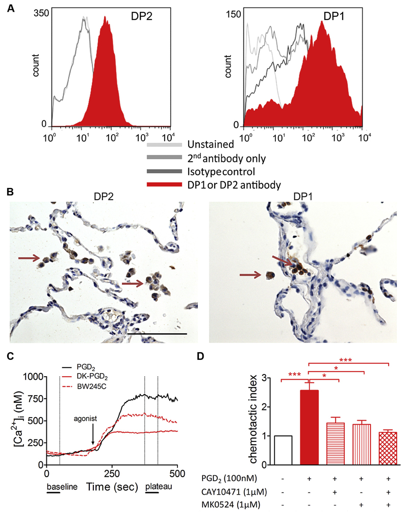
PGD2 receptors DP1 and DP2 are expressed on macrophages and induce Ca2+ flux and migration. A, Flow cytometric histograms of DP2 and DP1 staining (filled histograms) on MDMs, respectively. B, Immunohistochemistry of healthy human lung tissue showing DP2- and DP1-positive alveolar macrophages (arrows). C, Representative Ca2+ responses of MDMs over time. D, MDM migration toward PGD2 is blocked by DP1- and DP2-specific antagonists (n = 4-5). *P < .05 and ***P < .001.

