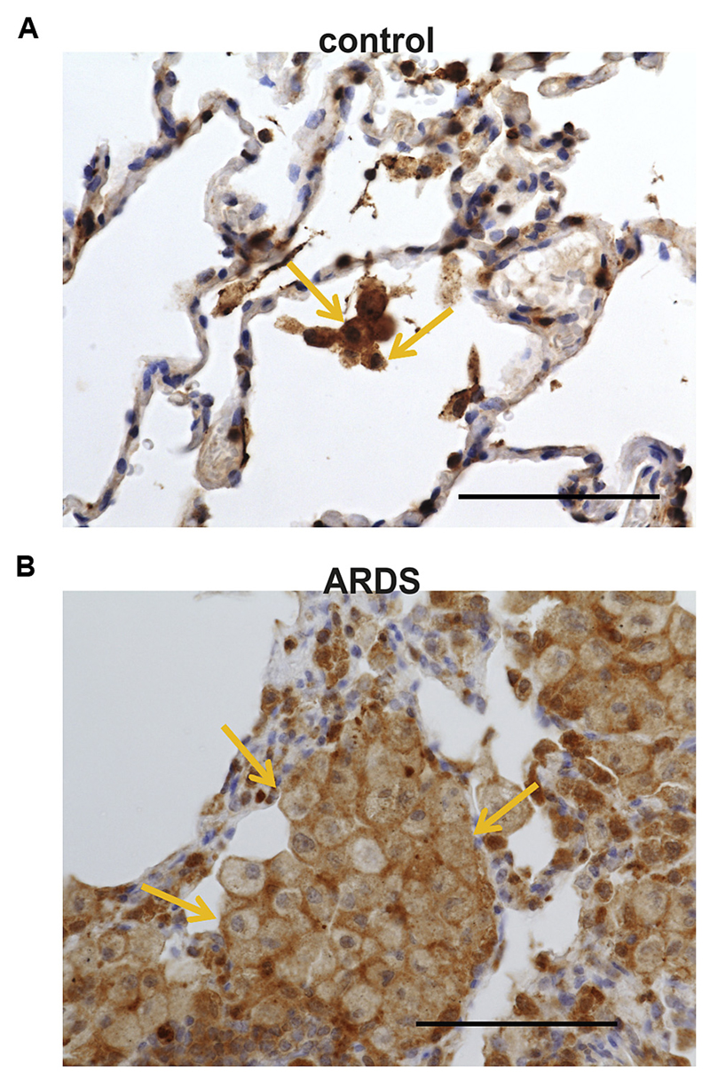
Fig 6.
Increased numbers of HPGDS-expressing cells in lungs of patients with ARDS. Representative immunohistochemical staining of human lung samples showing positive cells for HPGDS (brown) in a control subject (A) and a patient with ARDS (B). Stainings are representative pictures of 5 patients and control subjects. Note the high amount of HPGDS found in alveolar macrophages. Bar = 100 µm.

