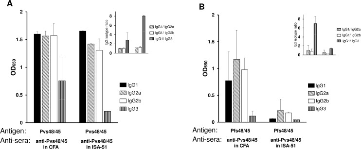
Fig 3. Analysis of antibody isotypes.
Anti-Pvs48/45 sera showing positive reactivity to Pfs48/45 in ELISA and Western blotting (4 out of 5 from CFA group and 2 out of 5 from Montanide ISA-51 group) were individually tested to compare immunoglobulin isotypes. ELISA plates coated with Pvs48/45 (panel A) or Pfs48/45 (panel B) were incubated with sera (1:10,000 dilution for Fig 3A and 1:100 dilution for Fig 3B). The plates were then incubated with peroxidase-conjugated goat anti-mouse IgG1, IgG2a, IgG2b and IgG3 (1:2,500 dilution) and processed as in standard ELISA. Shown are mean absorbance values for each isotype and the insets in panels A and B show relative proportions of IgG2a, IgG2b and IgG3 isotypes compared to IgG1. The error bars indicate SD.

