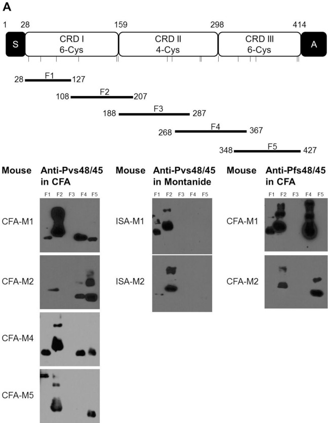
Fig 5. Reactivity of anti-Pvs48/45 sera to recombinant sub-fragments of Pfs48/45.
(A) Schematic representation of Pfs48/45 and the amino acid boundaries of five sub-fragments (F1, F2, F3, F4 and F5) of Pfs48/45. S, secretory signal sequence; A, anchor sequence; CRD, cysteine-rich domain. Bars show the relative positions of cysteine residues. (B) Western blotting analysis of the six anti-Pvs48/45 sera from CFA (CFA-M1, CFA-M2, CFA-M4 and CFA-M5) and Montanide ISA-51 (ISA-M1, ISA-M2) adjuvant groups with five overlapping fragments of Pfs48/45. All the anti-Pvs48/45 sera were tested at 1:1,000 dilution. The two anti-Pfs48/45 sera (CFA-M1, CFA-M2) were employed as positive control at a dilution of 1:10,000.

