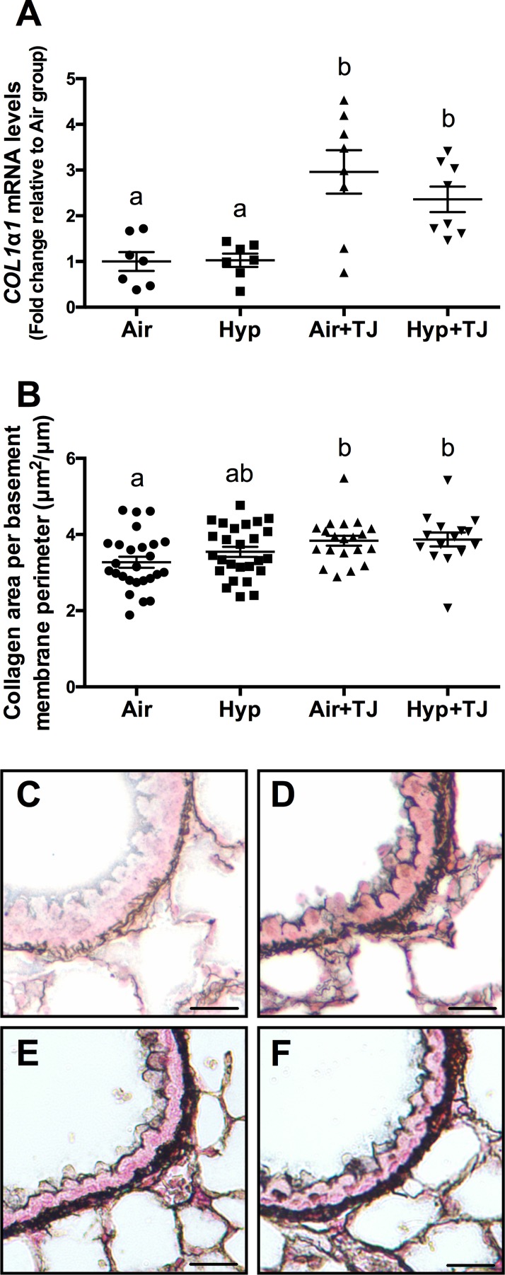
Fig 6. Collagen expression in the lung at P7d and P56d.
The mRNA expression of COL1α1 in lung tissue at P7d was significantly greater in the TJ groups compared to the non-TJ groups (A). The area of collagen in the outer bronchiolar wall at P56d was significantly greater in the TJ groups compared to the Air group (B). Data points represent values from individual animals and values with different letters are significantly different from each other (p<0.05). Representative images of lung sections stained for collagen (black staining) are representative of the bronchioles analyzed at P56d (C, Air; D, Hyp; E, Air+TJ; F, Hyp+TJ). Scale bar = 10μm.

