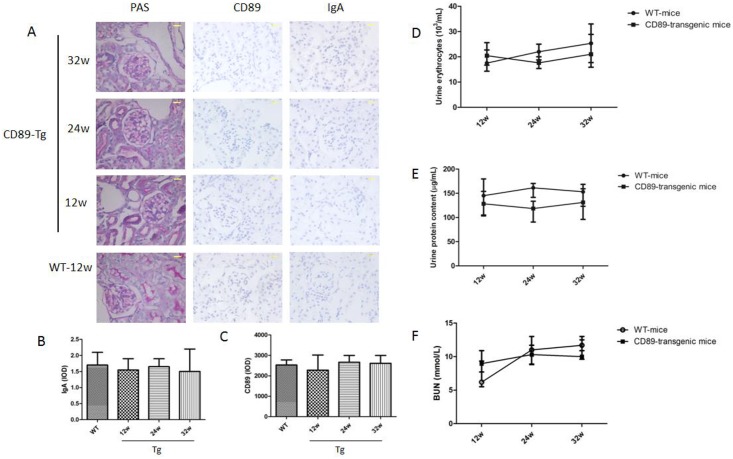
Fig 5. CD89 transgenic mice do not develop IgAN.
(A) From the left to the right: histology of kidney sections after periodic acid-Schiff (PAS), IgA and CD89 staining of kidneys from CD89 Tg mice (10 mice per group) at different ages. No mesangial IgA deposits or periglomerular CD89 cells were seen in kidneys from CD89 Tg mice; Bar: 25μm. (B and C) The IgA and CD89 IOD were quantified in 20 randomly chosen fields for each mouse at 400 magnification. n = 4 mice per group; (D and E) Hematuria (×1000 red cells/mL; D) and urine protein (μg/mL) were measured in the urine of 12 to 32-wk-old WT, CD89 Tg mice (7–14 mice per group). No difference was detected between WT and CD89 Tg mice. (F) BUN levels in CD89 Tg and WT mice.

