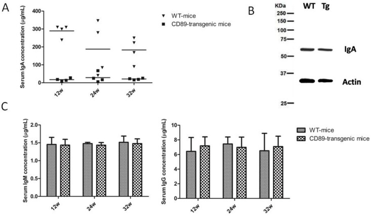
Fig 6. Serum IgA levels in CD89 transgenic mice were substantially decreased while IgA production by B lymphocytes was unaffected.
(A) Decreased serum IgA concentration in CD89 C57BL/6-Tg mice compared with WT mice. Serum from 12-, 24-, and 32-wk-old CD89 Tg mice (n = 4) and their littermate controls (WT; n = 4) was tested with a sandwich ELISA using a commercial mouse IgA detection kit. (B) There was no significant difference between the IgA expression of blood lymphocytes in CD89 C57BL/6-Tg mice compared with WT mice. Blood lymphocytes from 24-wk-old CD89 Tg mice and their littermate controls (WT) were isolated and lysed, and analyzed using Western blot. (C-D) Serum IgM (C) and IgG (D) concentrations from CD89 C57BL/6-Tg mice were unaffected when compared with WT mice. Serum from 12-, 24-, and 32-wk-old CD89 Tg mice (n = 4) and their littermate controls (WT, n = 4) was tested with a sandwich ELISA using mouse commercial IgM and IgG detection kits.

