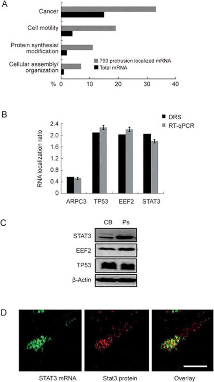
Figure 2.
Transcriptome analysis of purified mRNA from HCCLM3 protrusions. (A) Annotation analysis of the 793 HCCLM3 protrusion localized mRNAs compared to the total cellular mRNA detected at ≥5 RPKM. Annotations were made using the IPA Ingenuity platform. (B) RT-qPCR confirmation analysis of selected HCCLM3 protrusion localized RNA identified by DRS. RNA localization ratios are normalized to ARPC3 mRNA. (C) Western blot analysis of putative protrusion-localized proteins. Western blotting was performed on pooled material from three independent Boyden chamber experiments representing cell body fraction (CB) and protrusions (Ps). Subsequent Western blot analyses were performed with antibodies against Stat3, EEF2 and TP53. β-Actin was used as loading control. (D) Fluorescent in situ hybridization on STAT3 mRNA (left panel) and immunofluorescence (IF) on Stat3 protein (middle panel) in HCCLM3 cells. Scale bar: 2 μm.

