Figure 1.
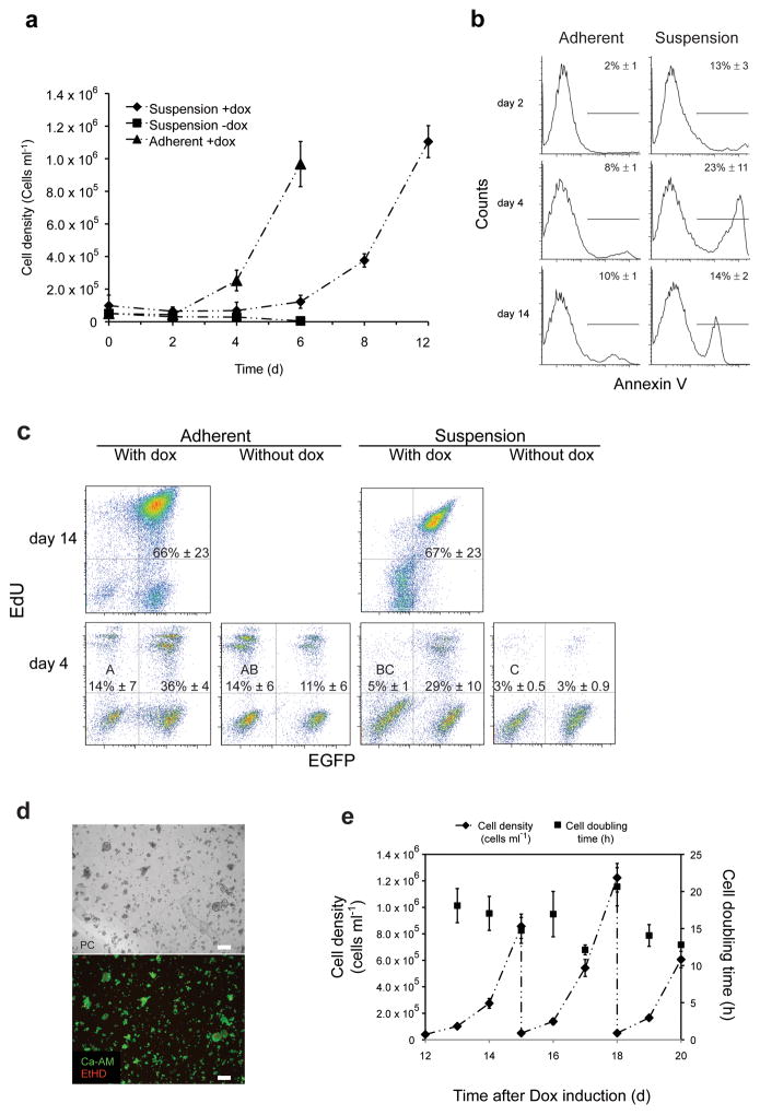
Secondary fibroblasts inducibly expressing reprogramming factors survive and proliferate in suspension. (a) Growth kinetics of inducible secondary fibroblasts cultured under the indicated conditions. Error bars s.d. (n = 3). (b) Fluorescence-activated cell sorting (FACS) analysis of AnnexinV surface localization for secondary fibroblasts cultured under the indicated conditions. Values are means ± s.d. (n ≥ 3). (c) FACS analysis of 5-ethynyl-2′-deoxyuridine (EdU) incorporation by secondary fibroblasts cultured under the indicated conditions. Percentages of GFP−/EdU+ populations that do not share a letter are significantly different (ANOVA, P = 0.003; and Tukey post-hoc with P = 0.0113) Values are means ± s.d. (n = 5). (d) Live/dead staining of secondary fibroblasts cultured in suspension in the presence of doxycycline at day six of culture. PC, phase contrast; red, ethidium homodimer-1 (dead); green, Calcein acetoxymethyl ester (live). Scale bars: 100 μm (e) Growth kinetics of doxycycline-induced secondary fibroblasts growing in mouse embryonic stem cell (mESC) medium from day 12–20. Suspension cultures inoculated with d12 cells (5 × 10⁴cells ml−1) were serially expanded with 20-fold media dilution every three days. Error bars s.d. (n = 3).

