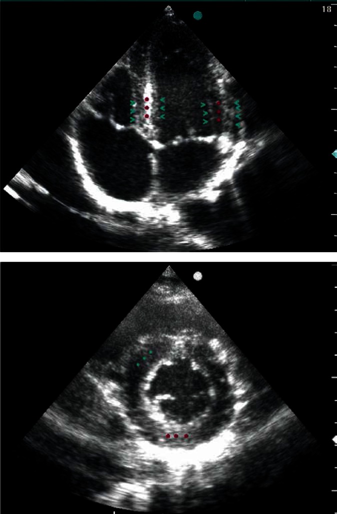
Fig. 2.
Two-dimensional four-chamber view (A) and a cross section of the left ventricle at the level of the papillary muscles (B), reflecting LV muscle fiber arrangement. Circumferentially oriented muscle fibers (perpendicular to the ultrasonic beam), located in the middle layer, reflect the ultrasonic waves more intensely than the oblique fibers, located in the external LV wall layers, which is expressed in the formation of intense, linear echo in the middle layer (red stars) and weaker echogenicity of the subendocardial and subepicardial layers (green arrow heads)

