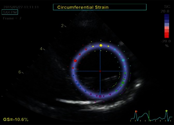
Fig. 3.
Parasternal transverse view of the left ventricle at the level of the papillary muscles. The image is used to determine the circumferential strain. The strain is analyzed in the tangential direction to the lines delineating the borders of the ROI (arrows) laid on the image in a similar manner as in Fig. 2. Similarly, the ROI was automatically divided into six equal color-coed segments corresponding to anatomical segments. The image was captured at late diastole, at a time point when speckles move away from each other, therefore the whole analyzed region is coded in a uniform blue color, indicating a homogeneous contraction throughout all analyzed segments

