. 2016 Jul 8;59(4):400–404. doi: 10.3340/jkns.2016.59.4.400

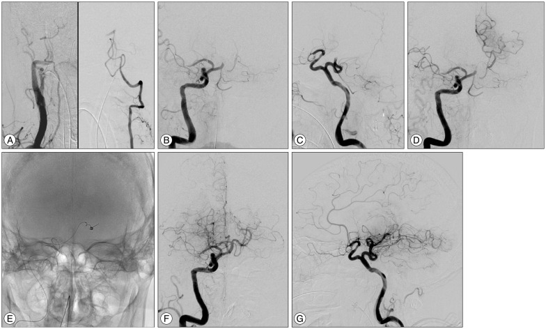
Fig. 2. A: Right innominate angiography showing a relatively hypoplastic right VA (left). Left vertebral angiography showing occlusion of the BA (right). The intracranial left VA is relatively hypoplastic. B and C: Right carotid angiography showing the occluded right middle cerebral artery and a persistent primitive trigeminal artery arising from the precavernous portion of the right internal carotid artery. D: Simultaneous angiography from the guiding catheter and the microcatheter indicating the location of the clot. E: Radioscopic image showing the Merci Retriever inserted into the left posterior cerebral artery. F and G: Final angiography after the endovascular treatment showing recanalization of the BA. VA: vertebral artery, BA: basilar artery.

Fig. 2