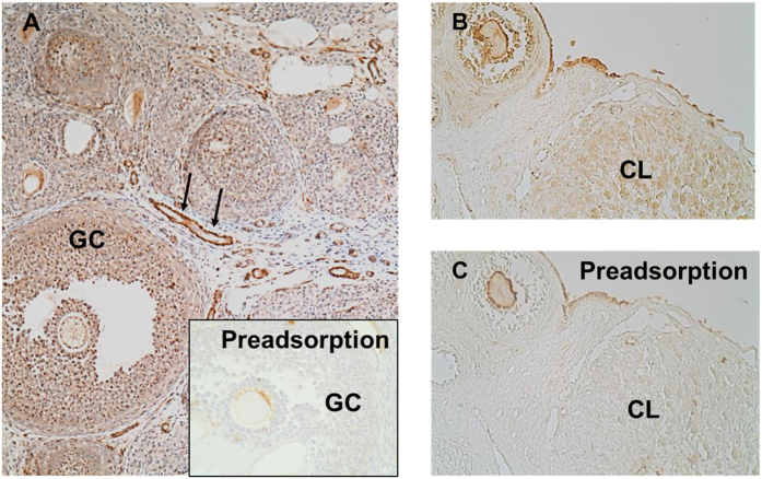
Figure 2. Immunohistochemical detection of AChE in rat ovary.
(A) GCs of preantral and antral follicles are positive for AChE in an immunohistochemical staining. AChE was also associated with blood vessels (arrows). Preadsorption of the antibody nearly abolished the staining (insert). (B) GCs, corpus luteum and surface epithelium show staining for AChE. (C) The staining nearly disappeared in the preadsorption control.

