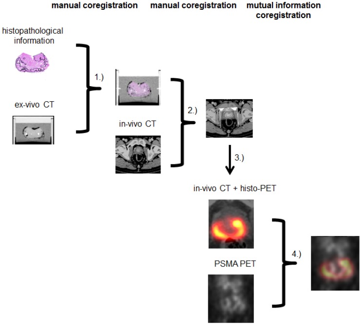
Figure 1.
Coregistration between step sections and ex-vivo CT was done by CZ in MITK (1). Ex-vivo CT (including histopathology) was matched to in-vivo CT by CZ and AC in MITK (2). Histopathological information was interpolated, binarized and smoothed to create histo-PET (3). Using rigid mutual information histo-PET and PSMA PET were coregistrated in PMOD (4).

