
Fig. 1.
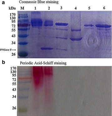
SDS-PAGE analysis of the purified recombinant β-glucosidase Bgl3A with and without deglycosylation treatment. a Coomassie Blue staining. Lanes M, the standard protein molecular weight markers; 1 the Bgl3A produced in P. pastoris; 2 the Bgl3A produced in P. pastoris and treated with PNGase F; 3 the Bgl3A produced in P. pastoris and treated with PNGase F followed by α-mannosidase; 4 the α-mannosidase; 5 the purified Bgl3A after sequential deglycosylation by PNGase F and α-mannosidase; 6 the Bgl3A produced in E. coli. b Periodic Acid-Schiff staining