Fig. 1.

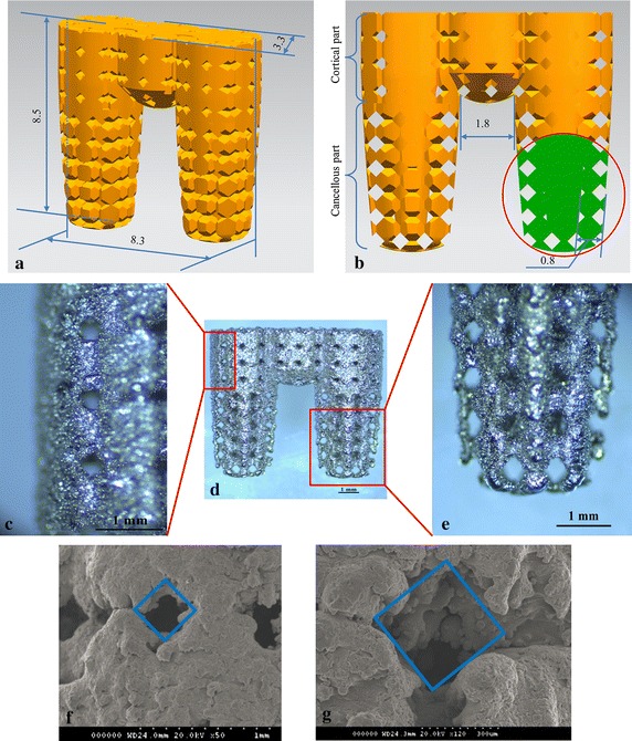
Multi-rooted implant (MRI). a Overall implant dimensions. b Partial cross-section of the MRI, illustrating the pore structure in detail. c The surface of the cortical bone region of fabricated MRI. d The overall profile of the fabricated MRI. e The surface of the cancellous bone region of the fabricated MRI. f Scanning electron microscopy (SEM) image of the cortical bone region of the implant; the pore structure width was approximately 290 µm. g SEM image of the cancellous bone region; the pore structure width was approximately 390 µm
