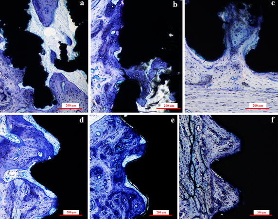
Fig. 3.

Histological sections of the MRIs and RBM implants. Representative sections of the MRIs in rabbit hind limbs at a 4 weeks, b 8 weeks, and c 12 weeks after implantation, and RBM implants in rabbit hind limbs at d 4 weeks, e 8 weeks, and f 12 weeks after implantation. The sections were stained with toluidine blue
