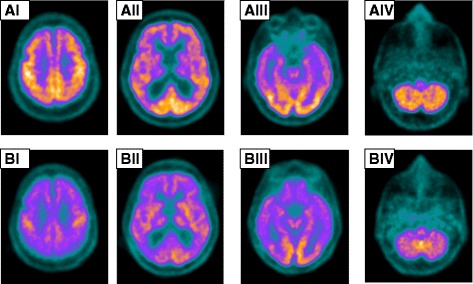
Fig. 2.

Cerebral scans (axial slices, I–IV) obtained by 2-deoxy-2-[fluorine-18]fluoro-D-glucose integrated with computed tomography-positron emission tomography. Cerebral 2-deoxy-2-[fluorine-18]fluoro-D-glucose integrated with computed tomography-positron emission tomography was performed 23 months before the patient’s death. a Four representative axial slices (AI–AIV) show decreased fluorodeoxyglucose uptake in the frontal and temporal lobes and normal uptake in the posterior cingulum and occipital cortex. Note that the hypometabolism in the affected regions is greater in the right hemisphere. b Cerebral 2-deoxy-2-[fluorine-18]fluoro-D-glucose integrated with computed tomography-positron emission tomography performed 3 months before the patient’s death shows the general reduction in radiotracer uptake in the same axial levels (BI–BIV)
