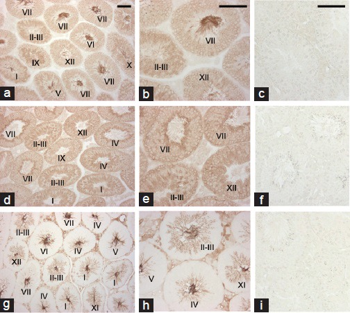
Figure 3.

Immunohistochemical localization of basigin, MCT1 and MCT2 in mouse testes. Testes from adult mice were immunostained with anti-basigin (a and b), anti-MCT1 (d and e) or anti-MCT2 (g and h) antibody. Roman numerals indicate the stage of the seminiferous epithelium. (c, f, i) negative controls lacking primary antibodies, anti-basigin (c), anti-MCT1 (f) and anti-MCT2 (i). Scale bars: 100 μm.
