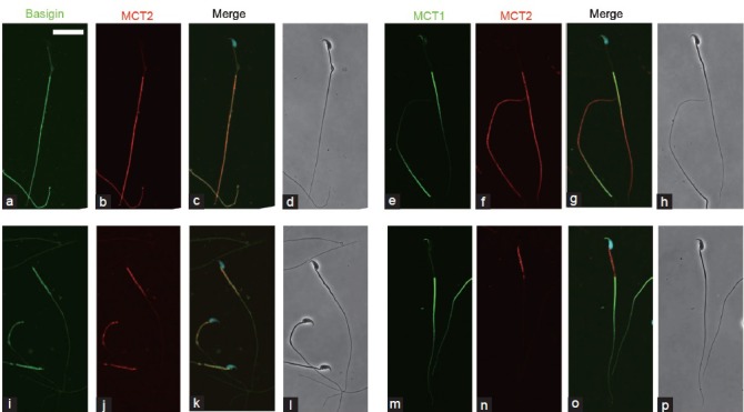
Figure 5.

Indirect immunofluorescence localization of basigin, MCT1, and MCT2 in spermatozoa. Spermatozoa from the caput (a–h) and cauda (i–p) epididymis were immunostained with anti-basigin and anti-MCT2 antibodies (a–d, i–l) as well as with anti-MCT1 and anti-MCT2 antibodies (e–h, m–p). Immunoreaction for basigin (a and i) and MCT1 (e and m) is represented in green, and that for MCT2 (b, f, j, n) in red. (c, g, k, o) merged images with green, red and Hoechst staining (blue). More than 90% of spermatozoa showed similar immunoreactions. (d, h, l, p) phase contrast images. Scale bar: 20 μm.
