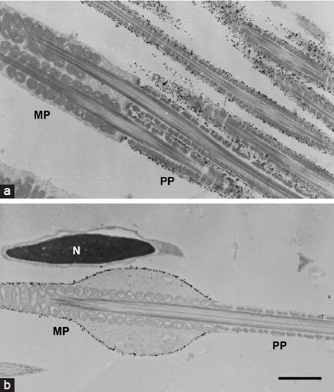
Figure 6.

Immunoelectron micrographs of MCT2 in spermatozoa. Spermatozoa from the caput epididymis (a) and ductus deferens (b) were immunolabeled with anti-MCT2 antibody. The majority of gold particles were seen on the surface of the principal piece (PP), not on the midpiece (MP) of the sperm flagella from the caput epididymis (a). In the sperm from ductus deferens, the immunoparticles were detected on the MP, not on the PP (b). N: nucleus of the sperm head. Scale bar: 1 μm.
