
Figure 3.
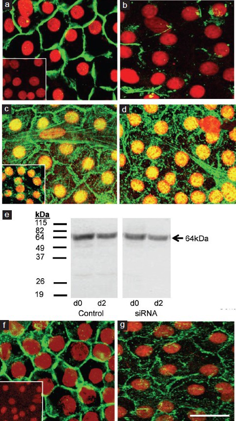
Effect of claudin-11 siRNA on the localization of occludin to the TJ and β-catenin to the adherens junction, and occludin protein expression. Cultured immature rat Sertoli cells were fixed after 2 days of claudin-11 siRNA treatment. Immunocytochemical analysis was conducted on claudin-11 (green) localization around the periphery of Sertoli cells (nuclei in red) at the TJ in cells which received medium plus transfection reagent (panel a) or Clau-11-A/B siRNA treatment (panel b). Staining for occludin (green) at the Sertoli cell TJ was also conducted in medium plus transfection reagent (panel c) and Clau-11-A/B siRNA treated cells (panel d). To test for any effects of claudin-11 siRNA on other junctional types, β-catenin (green) of the adherens junction was also stained in medium plus transfection reagent (panel f) and siRNA (panel g) treated cells. Western blot analysis for occludin (panel e; arrow) was conducted on cell lysates on the day of Clau-11-A/B siRNA addition (indicated as d0) and after 2 days of treatment (indicated as d2). Note the panel on the left represents control extracts from the same time-point which did not receive siRNA. Molecular weight marker in kDa is provided to the left. For immunocytochemistry, bar = 50 μm. Inset = negative controls for claudin-11, occludin and β-catenin.