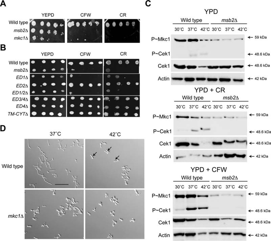
Fig. 4. Msb2 regulates the PKC pathway.
A. Growth of strains in YPD, YPD + CFW (100 µg/ml) and YPD + CR (100 µg/ml). Serial dilutions were spotted, and plates were incubated for 48 h at 37°C.
B. Growth of Msb2 deletion mutants was examined as in panel 4A.
C. Activity of the CEK and PKC pathways in response to different stimuli. Mid-log phase cells in YPD were incubated in YPD media alone, YPD + CFW (100 µg/ml) and YPD + CR (100 µg/ml) for 3 h at the indicated temperatures. Total protein (20 µg) was examined by immunoblot analysis with α-phospho p42/44 MAPK rabbit monoclonal antibodies, α-Cek1 and α-Act1 as a control for total protein levels.
D. Cells were grown to mid-log phase in YNB + 2% Glucose for 3 h at 37°C and 42°C and observed by microscopy at 20X magnification. Arrows, examples of hyphae at 42°C. Bar, 10 microns.

