Figure 1.

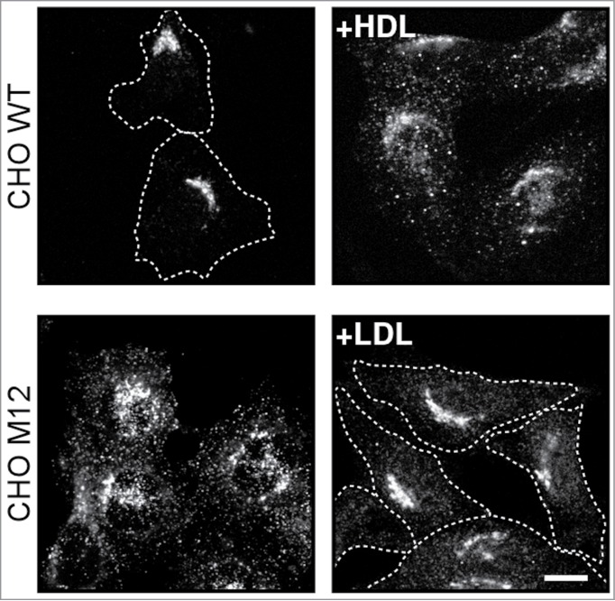
Low and High Density lipoproteins differentially regulate Stx6 localization. (A) Chinese Hamster Ovary (CHO) wild-type (wt) and Niemann Pick Type C (NPC1) mutant CHO M12 cells were incubated ± High Density lipoproteins (HDL; 0.1 mg/ml) for 30 min or Low Density lipoproteins (LDL; 0.05 mg/ml) for 24 hours as indicated. Cells were fixed and immunolabeled with anti-Stx6. Dotted lines indicate cell shape. Bar is 10 μm. Upper panels: Control CHOwt cells show compact staining of Stx6 in the Golgi. HDL induces partially scattered Stx6 staining, resembling recycling endosomes. Lower panels: NPC1 mutant CHO M12 cells show dispersed, endosomal Stx6 staining. Prolonged LDL loading re-establishes compact Stx6 staining in the Golgi.6
