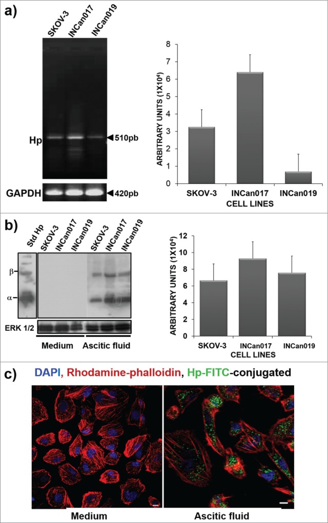
Figure 1.

Haptoglobin expression in the SKOV-3 ovarian cancer cell line and primary culture cells (INCan017 and INCan019). (A) RT-PCR assays using specific primers for Hp, showing an amplification product corresponding to 510 bp; (B) Western blot (C) and confocal microscopy (C) analyses using an anti-haptoglobin monoclonal antibody (1:1000, and 1:100, respectively) of cells that were harvested from culture medium or ascitic fluid. The cells in culture medium did not express the protein, whereas these cells, when stimulated with ascitic fluid from an ovarian cancer patient, expressed the haptoglobin protein. Bar scale = 100 μm.
