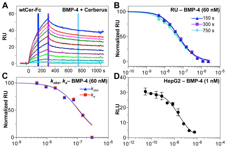
Figure 4. Inhibition of BMP-4 – Cerberus binding with Cerberus.
(A) Cerberus prevents BMP-4 – Cerberus-Fc binding. Cer-Fc was captured on the sensor chip and 60 nM BMP-4 preincubated with 0 nM (blue), 4.6875 nM (red), 9.375 nM (magenta), 18.75 nM (green), 37.5 nM (maroon), 75.0 nM (dark blue), 150.0 nM (purple), 300.0 nM (bright green), 600.0 nM (teal), 1200.0 nM (cyan), and 2400.0 nM (grey), Fc free Cerberus was injected over the sensor chip. (B) Background subtracted raw RU values were taken for each Cerberus concentration at 150 seconds (blue), 300 seconds (purple) and 750 seconds (cyan). RU values for each group were normalized and fitted using non-linear regression in GraphPad. SEs are similar to those observed for ActRIIA-Fc inhibition in 2D, but are omitted here for clarity. (C) Analysis of kobs (blue) and ka (red) data for Cerberus-Fc. kobs and ka values were obtained by individually fitting each injection curve. kobs and ka values were normalized and fitted using non-linear regression in GraphPad. SEs are similar to those observed for ka analysis in 2F, but are omitted here for clarity. (D) BMP-4 mediated Smad1/5/8 signaling and inhibition with Cerberus. HepG2 cells were transfected with SMAD1/5/8 responsive reporter and control plasmids. Cells were treated with 1.0 nM BMP-4, and Cer-Fc at the following concentrations: 0.0 nM, 0.03 nM, 0.09 nM, 0.27 nM, 0.82 nM, 2.47 nM, 7.41 nM, 22.2 nM, 66.6 nM, 200 nM, and 600 nM. Concentrations in panels B–D are shown in logarithmic scale.

