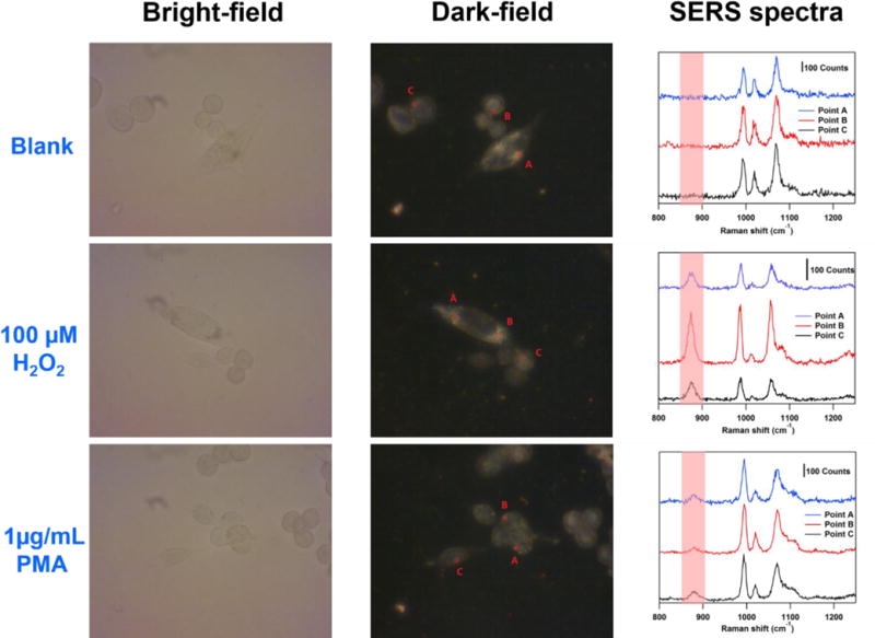
Figure 8.

Bright-field images (left column) and dark-field microscopy images (middle column) of SW620 cells incubated with the nanoprobe after 24 hours respectively. Right column: SERS spectra obtained from the points denoted in the corresponding dark-field microscopy images in the same row. Samples at the first row are blank. Samples at the second row and the third row are treated with 100 μM H2O2 and 1 μg/mL PMA respectively for 1 hour. (λex = 785 nm, Plaser = 1.3 mW, t = 5 s).
