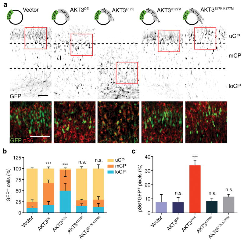
Figure 3. AKT3 kinase activity is essential for aberrant migration phenotype.
(a) In utero electroporation at E14.5 of GFP vector encoding AKT3 variants, harvested at E18.5. Images were inverted for visibility, co-stained with phospho-S6 (pS6) of boxed regions below, enhanced contrast for visibility. (b) Localization of GFP+ cells quantified in upper (uCP); middle (mCP), and lower cortical plate (loCP). (c) Quantification of pS6+GFP+ pixels for the images described in (a). Values: mean ± s.d. (n = 3, 3, 6, 5 and 5 for each condition). n.s., not significant; ***, P < 0.001, G-test of goodness-of-fit (b); Student’s t-test (c). Scale bars: 100 μm.

