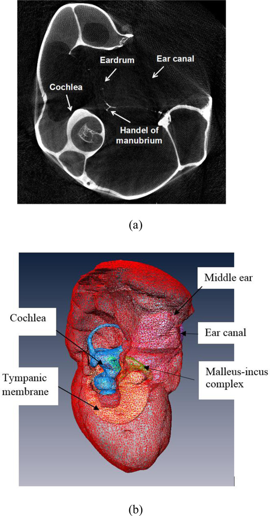
Figure 1.
A typical µCT image and the 3D geometrical model of a chinchilla left ear. a A µCT image of the chinchilla bulla, showing a portion of the outer ear canal, eardrum, manubrium, middle ear cavity and cochlea. b Lateral view of the 3D model of chinchilla middle ear and cochlea reconstructed from µCT images.

