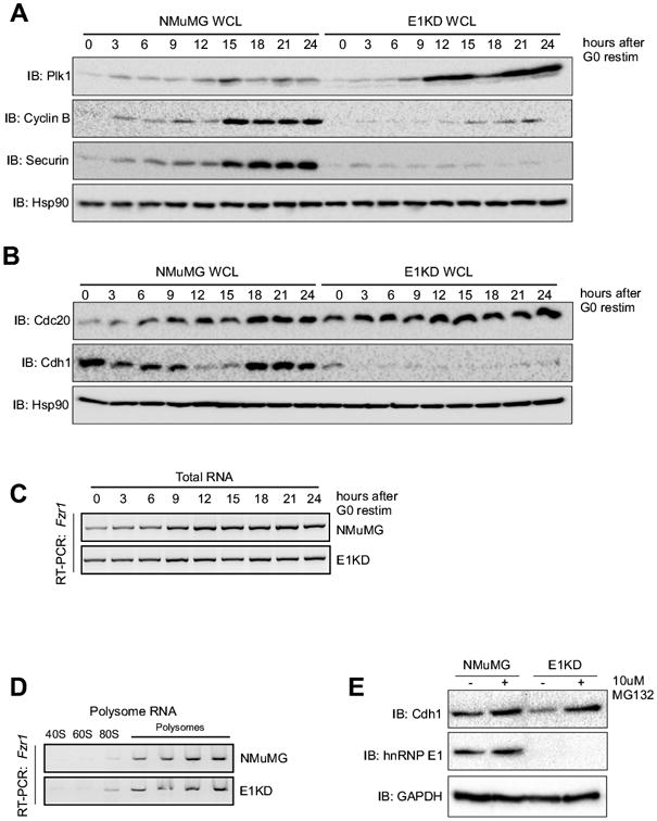
Figure 4. APC/C-Cdh1 function is lost in E1KDs, as well as expression of Cdh1.

(A) Immunoblot analysis of protein expression of Plk1, cyclin B1, securin, and Hsp90 (control) using whole cell lysates isolated from G0-synchronized and released NMuMG and E1KD cells (B) Immunoblot analysis of protein expression of Cdc20, Cdh1, and Hsp90 (control) using whole cell lysates isolated from G0-synchronized and released NMuMG and E1KD cells. (C) Semi-quantitative RT-PCR analysis using gene specific primers for Fzr1 on total RNA isolated from NMuMG and E1KD cells synchronized as described in A above. (D) Semi-quantitative RT-PCR analysis with gene specific primers for Fzr1 using RNA isolated from polysome profiling of NMuMG and E1KD cytosolic extracts. (E) Immunoblot analysis of protein expression in whole cell extracts prepared form NMuMG and E1KD cells treated ± 10 μM MG132 for 1 hour using α-Cdh1, α-hnRNP E1, and α-GAPDH (control) antibodies.
