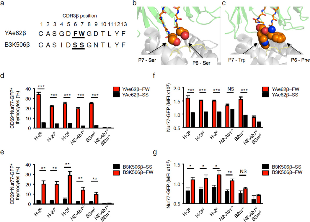
Figure 3.
B3K506β+ pre-selection thymocytes expressing the CDR3β P6–7 doublet, FW, show increased recognition of self-pMHC ligands. (a) Alignment of CDR3β sequences from Yae62 and B3K506 TCRs, with residues P6 and P7 sequences in bold. (b,c) The YAe62 (b) and B3K506 (c) CDR3β residues P6 and P7 (orange) interact with amino acids of the peptide (yellow) and MHCII (white), PDB 3C6L, 3C5Z. (d,e) Quantification of the frequency at which pre-selection thymocytes express CD69 and Nur77-GFP following culture with BM-DC. (f,g) Nur77-GFP expression levels on activated Nur77+CD69+ pre-selection thymocytes from TCRβ retrogenic mice expressing the YAe62β chain (d,f) or the B3K506β chain (e,g) carrying the CDR3β P6–7 doublet, FW or SS, following incubation with BM-DC. BM-DC were derived from C57BL/6 (H2b), NOD (H2g7), Balb/c (H2d), C57BL/6.H2-Ab1−/−, C57BL/6.B2m−/−, or C57BL/6.H2-Ab1−/−B2m−/− mice. NS, not statistically significant, *P <0.05; **P < 0.01; ***P < 0.001; ****P < 0.0001 (unpaired two-tailed Student’s t-test). Data are combined from two independent experiments YAe62β-FW (n = 8), YAe62β-SS (n = 10), B3K506β-SS (n = 4), B3K506β-FW (n = 7) retrogenic mice were analyzed four weeks post-reconstitution (d–g, mean ± s.e.m.).

