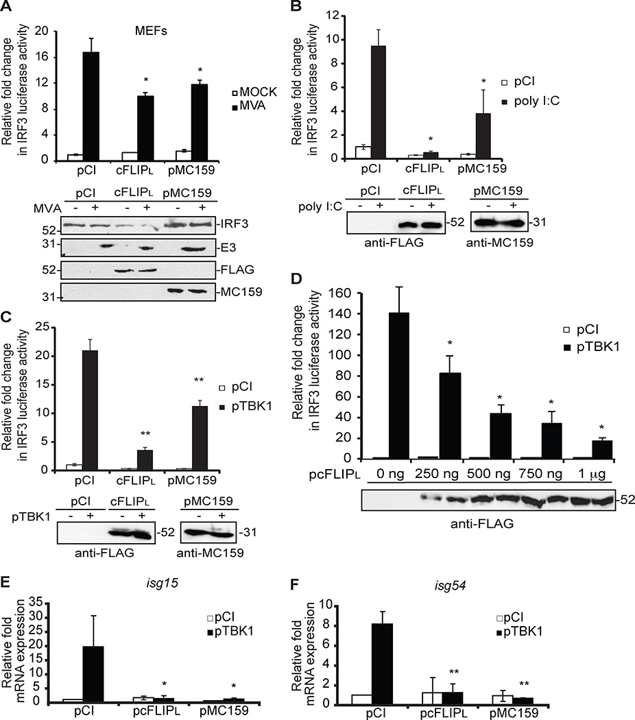
Figure 1. cFLIPL inhibits IRF3-controlled luciferase activity.
Luciferase activity in (A) MEF or (B) 293T-TLR3 or (C–D) 293T cells transiently co-transfected for 24 hours with luciferase reporter plasmids (pIRF3-luc, pRL-null) and 1000 ng control plasmid (pCI) or plasmids encoding cFLIPL or MC159. (A) Cells were either mock-infected or infected with MVA (MOI = 5 PFU/cell) or (B) incubated with 1000 ng of poly I:C or (C and D) co-transfected with pTBK1. Cells were lysed at (A and B) 6 h or (C and D) 24 h later. Results are shown as fold-induction of luciferase activity, relative to those of pCI-transfected cells. Immunoblot analysis of lysates also was performed. (E and F) Quantitative RT-PCR (qPCR) analysis of (E) isg15 and (F) isg54 mRNA from 293T cells co-transfected with pTBK1 and 1000 ng control plasmid (pCI) or plasmids encoding cFLIPL or MC159 for 24 h. Results are presented as isg15 and isg54 mRNA relative to β-actin mRNA expression for each sample, and recorded as relative to those of cells transfected with empty vector (pCI). *P < 0.05, compared with cells transfected with empty vector. All experiments were performed at least three times, and data are expressed as the mean +/− s.d.

