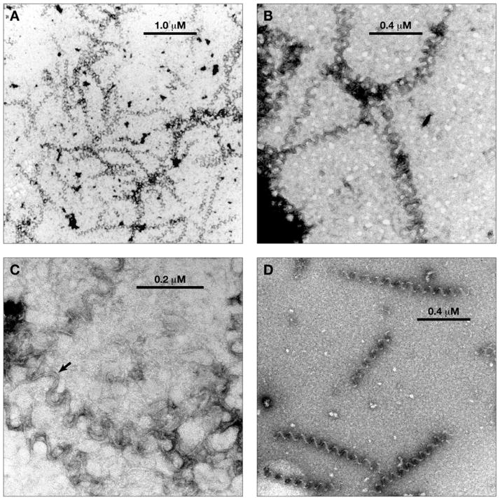
Fig. 3.
Electron micrographs of aberrant polymer formed with (−)-rhazinilam. Magnification is indicated by the bars in each panel. A. Lower magnification view. B. Middle magnification view. C. Higher magnification view. The arrow indicates a segment of polymer where a 2 filament substructure is clearly visible. D. Middle magnification view of polymer mildly fixed with glutaraldehyde (0.2%) before sample was applied to the grid.

