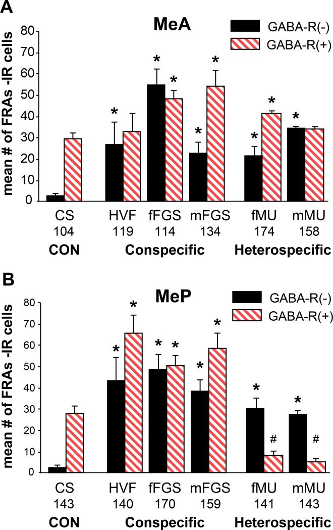
Figure 4. FRAs activation in cells that are GABA-Receptor (+/−) in Medial Amygdala.
A. MeA: GABA-R(−) cells in MeA were activated by all stimuli. GABA-R(+) cells were activated by all but HVF and mMU. mFGS and fMU activated more GABA-R(+) cells than GABA-R(−).
B. MeP: FRAs expression in both GABA-R(+) and GABA-R(−) cells in MeP was significantly different than with CS controls for all stimuli. However: heterospecific stimuli significantly suppressed GABA-R(+) cells (#; suggesting GABA inhibition) - and significantly activated GABA-R(−) cells in MeP. Conspecific stimuli activated both phenotypes. Mean total numbers of GABA-R(+) cells are shown for each area below the bars in the graph.

