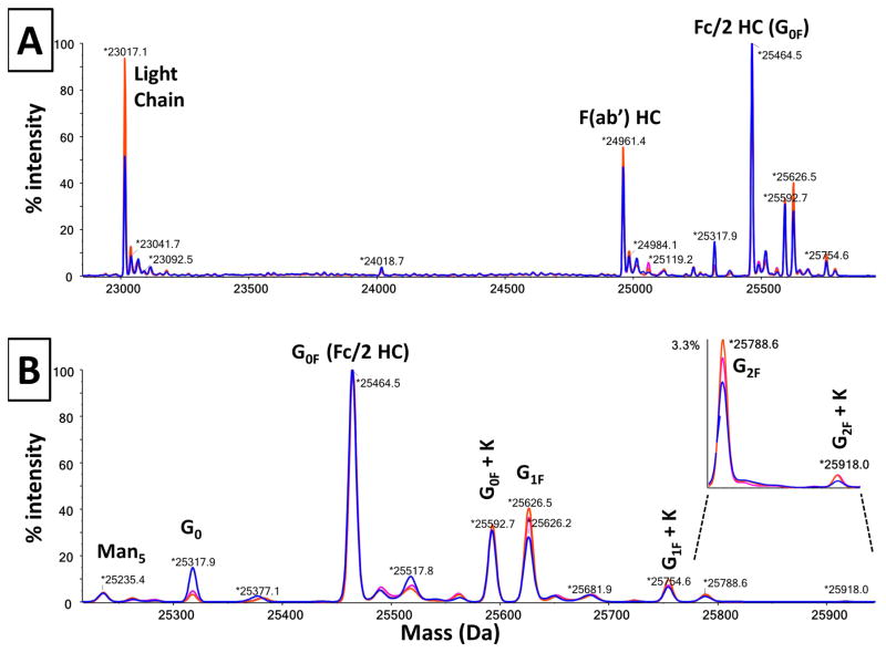
Figure 3. Overlay of 3 mass spectra obtained from 3 cell lines of the h2E2 anti-cocaine mAb.
Panel A shows the overlay of the 3 reconstructed mass profiles over the mass range covering all of the fragments. Panel B shows an overlay of the expanded portion of the mass spectra defining the glycoforms for the 3 cell lines of the h2E2 anti-cocaine mAb. Cell line 85-blue profile; Cell line 188-magenta profile; Cell line 323-orange profile.

