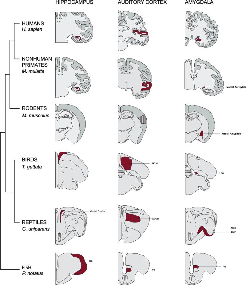
Figure 1. Aromatase is typically expressed in brain regions crucial for cognition among vertebrates.
Aromatase expression is abundantly expressed within the hippocampus, auditory cortex/forebrain, and amygdala of several representative species across a wide range of classes. Black stripped-filled brain regions indicate no reported presence of aromatase, whereas maroon-filled brain regions indicate detectable presence of aromatase as assessed through various techniques. Briefly, 1) hippocampus - humans: [22,23]; rhesus macaques: [24]; mice: not seen in hippocampus [19,20]; but see [25]; birds: [26,27]; reptiles (medial cortex): [28,29]; fish (dorsolateral telencephalon): [30,31]; 2) auditory cortex/forebrain – humans: [32,33]; rhesus macaques: [24]; birds (caudomedial nidopallium; NCM [34]): [26,27]; reptiles (anterior dorsal ventricular ridge; ADVR [34]): [28]; fish (posterior portion of the ventral telencephalon; Vp): [30,35,36]; 3) amygdala - humans: [37]; rhesus macaques: [38]; mice: [19]; birds (nucleus taenia; TnA): [27]; reptiles: [28,29,39,40]; fish (supracommissural nucleus of the ventral telencephalon; Vs [41,42]): [30,36].

