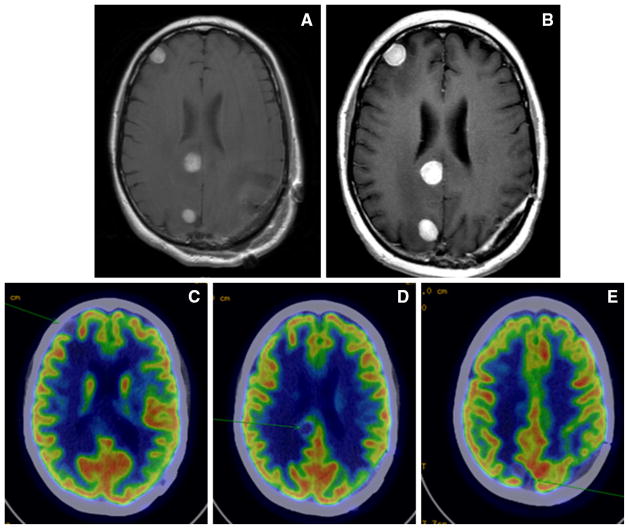
Fig. 2.
Neuroimaging from one patient who was classified as having progressive disease by modified World Health Organization criteria and stable disease by immune-related response criteria. a Contrast T1-weighted image pre-treatment. b Four months post-treatment showing 33 % growth of brain metastases. c–e Positron-emission tomography scan 4-months post-treatment showing that areas of growth on magnetic resonance imaging are photopenic and therefore not likely to represent viable disease

