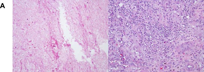
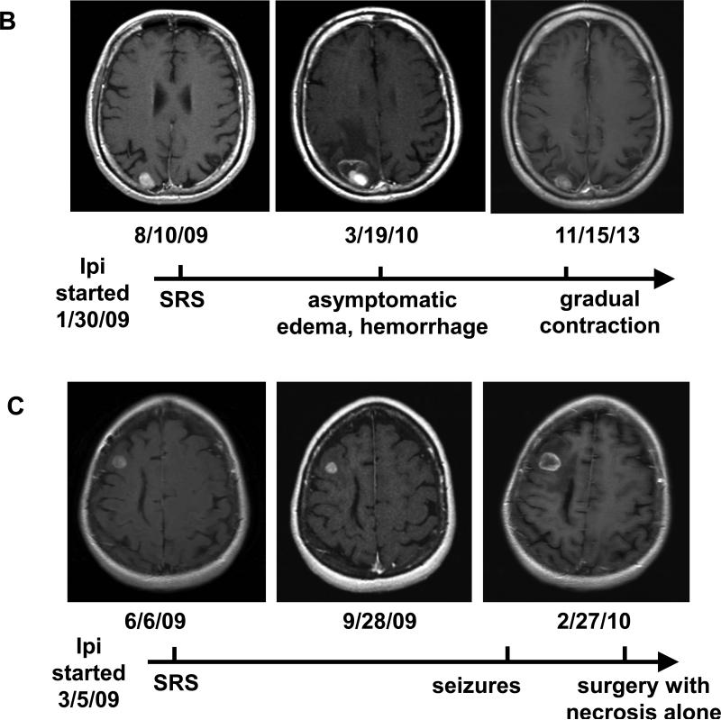
Figure 2.
Pathologic and radiologic findings after treatment with SRS during ipilimumab. (a) Micrographs at 200X of a brain lesion resected 5 months after treatment, demonstrating no viable tumor but (left) cellular effacement and granular eosinophilic debris characteristic of necrosis and (right) lymphocytic and histiocytic infiltration. (b) and (c) Brain MRI findings of two patients who received SRS during ipilimumab.


