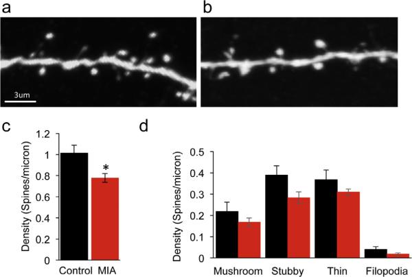
Fig. 1.
Reduced cortical dendritic spine density in young MIA offspring. Confocal images of layer 5 pyramidal neuron apical tuft dendrites from P17 offspring of control (a) and MIA (b) YFP-H mice. (c) MIA results in a reduction in total dendritic spine density (control: 1.014 ± 0.07 spines/micron, n = 4; MIA: 0.78 ± 0.04 spines/micron, n = 6, P = 0.018, unpaired t-test). (d) MIA results in a general reduction in density of all dendritic spine categories although these differences did not reach significance following multiple comparison corrections (mushroom P = 0.27, stubby P = 0.06, thin P = 0.185 and filopodia P = 0.11, multiple t-test with Sidak–Bonferroni correction. Data are represented as mean ± SEM.

