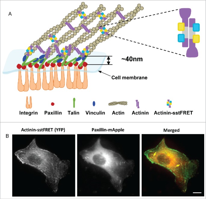
Figure 1.

Co-transfection of MDCK cell with actinin-sstFRET and paxillin-mApple. (A) Schematic of a focal adhesion complex, showing that the FRET force sensor (actinin-sstFRET) and paxillin-mApple are located in separate layers of the adhesion complex. (B) Actinin-sstFRET (YFP channel, green), paxillin-mApple (RFP channel, red) and merged images of a co-transfected MDCK cell. Yellow arrows indicate FAs at the ends of the stress fibers. Scale bar =10 μm.
