
Figure 1.
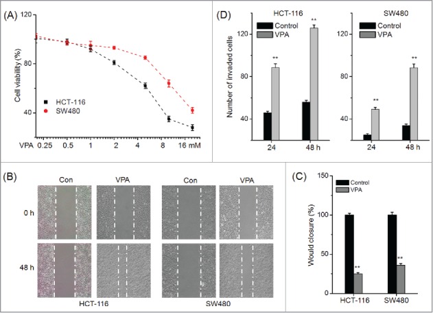
VPA promotes the in vitro motility of CRC cells. (A) HCT-116 and SW480 cells were treated with various concentrations of VPA for 48 h, and then the cell viability was assessed by use of MTT assay; (B) Representative images of wounds at 0 and 48 h in the presence or absence of 1 mM VPA; (C) Quantitative analysis of wound healing assay for CRC cells treated with 1 mM VPA for 48 h; (D) HCT-116 or SW480 cells were allowed to invade spread through the matrix gel and into the under-side of the filter for 24 h and 48 h in the presence or absence of 1 mM VPA. The number of invaded cells were fixed, stained, photographed, and compared with the control group. **p < 0.01 compared with control.