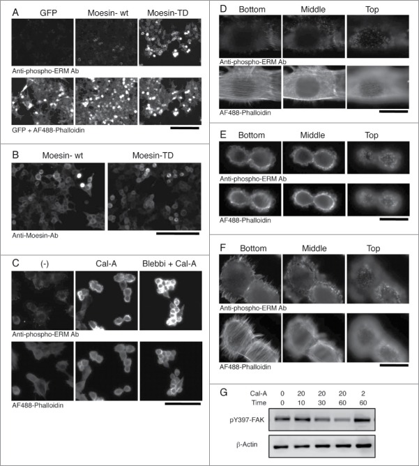
Figure 4.
Subcellular localization of phospho-ERM proteins. (A) Immunocytochemistry of HEK293T transfectants with anti-phospho-ERM Ab. Up: fluorescent images of anti-phospho-ERM Ab. Down: fluorescent images of GFP and AF488-labeled Phalloidin. Magnification: x200. Scale bar: 200 μm. (B) Immunocytochemistry of HEK293T transfectants with anti-Moesin Ab. Staining with anti-Moesin Ab was detected at the edge of cells in Moesin-TD transfectants, while detected rather at the cytoplasm in Moesin-wt transfectants. Magnification: x200. Scale bar: 200 μm. (C) Immunocytochemistry of Cal-A-treated NIH3T3 cells. NIH3T3 cells untreated (left), treated with 20 nM Cal-A for 20 min (middle), and treated with Cal-A after Blebbistatin (Blebbi) pretreatment (right) were analyzed with anti-phospho-ERM Ab (up) and AF488-labeled Phalloidin (down). Cal-A treatment augmented phospho-ERM, induced cell body retraction and bleb formation. Fewer blebs were observed in Blebbistatin-pretreated cells. Magnification: x200. Scale bar: 200 μm. (D) Non-treated NIH3T3 cells. (E, F) Cal-A treated NIH3T3 cells. From D to F: Up: anti-phospho-ERM Ab. Down: AF488-labeled Phalloidin. Magnification: x1000. Scale bar: 20 μm. Side (E, F) and top (E) parts of Cal-A-treated cells were covered with blebs. Phospho-ERM and Phalloidin staining were detected at the rim of blebs. (G) Immunoblotting with anti-pY397-FAK. NIH3T3 cell lysates used in Figure 1B were analyzed by immunoblotting with anti-phospho-FAK (Y397) and anti-β-Actin Abs. Phospho-Y397-FAK was decreased over time after Cal-A addition to 20 nM. Concentration of Cal-A: nM, time: min.

