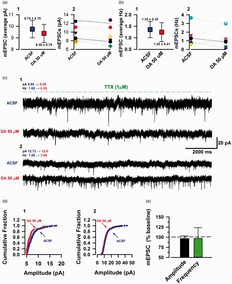
Figure 4.
DA can potentiate and inhibit mEPSC frequency. TTX sensitive mEPSC currents in the ACC layer II/III pyramidal neurons. (a) 1: Blue box indicating the mean value of raw amplitudes (pA) data at baseline (5 min). Red box: mean sEPSC amplitudes during DA (15–20 min post DA (50 μM) application) course of action. 2: Individual neuron amplitudes at baseline and DA (n = 9/6 mice). (b) 1: Average raw frequencies (Hz) at baseline (blue box) and DA (blue box). 2: Individual neuron frequencies at baseline and DA. (c) 1: Example mEPSC trace of a neuron with inhibited frequency by DA application. 2: Example mEPSC trace of a neuron with a very large increase in frequency in response to DA. (d) 1: Cumulative fraction of amplitudes for the neuron with inhibition of frequency (trace c, 1). 2: Cumulative fraction of amplitude for neuron with increase in frequency (trace c, 2). (e) DA had no significant influence on mEPSC amplitude (P = 0.738) and the mixture of potentiation and inhibition of frequency at the individual neuronal level yielded an insignificant statistical value (P = 0.842). Error bars represent SEM. ACSF: artificial cerebrospinal fluid; mEPSCs: miniature excitatory postsynaptic currents; DA: Dopamine.

