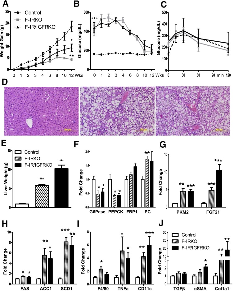
Figure 7.
HFD accelerates liver injury in lipodystrophic mice. A: Body weight of control, F-IRKO, and F-IR/IGFRKO mice assessed during 12 weeks of HFD feeding. B: Blood glucose levels in the same mice assessed at the indicated times. C: Glucose tolerance test of control, F-IRKO, and F-IR/IGFRKO mice after 12 weeks of the HFD. Results are mean ± SEM of 6 to 11 animals per group. D: H&E-stained liver sections from the same mice. One representative section from six mice per group is shown. E: Liver weight of control, F-IRKO, and F-IR/IGFRKO mice after 12 weeks of the HFD. mRNA expression of gluconeogenic enzymes (F) and the expression of Pkm2 and Fgf21 mRNA (G) after 12 weeks of the HFD. mRNA levels of genes involved in de novo lipogenesis (H), inflammation (I) and fibrosis (J) after 12 weeks of the HFD. Results are mean ± SEM of four to six animals per group. *P < 0.05, **P < 0.01, and ***P < 0.001 compared with controls.

