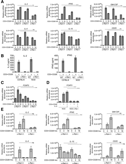
Figure 4.
c-Rel deficiency decreases cytokine and FOXP3 expression in NOD mice. A: CD4+ T cells were purified from c-Rel+/+, c-Rel+/− and c-Rel−/− NOD mice by magnetic-activated cell sorting. Cells were stimulated with plate-bound anti-CD3 and anti-CD28 antibodies (2 μg/mL each) for the indicated time points. Gene expression was analyzed by real-time PCR from at least three mice per genotype in triplicate. B: Analysis of the expression of IL-2 and IFN-γ in culture supernatant by ELISA. CD4+ T cells from C57BL/6 and NOD mice were treated as in A for 18 h. Data are the mean of triplicates from three mice per group. C: Expression of FOXP3 was analyzed as in A in triplicate from three mice per group. D: CD4+ T cells were treated as in A, and the effect of exogenous IL-2 (50 units/mL) on FOXP3 expression was analyzed. Data are the mean of triplicates from three mice per genotype. E: CD8+ T cells were purified from c-Rel+/+ and c-Rel−/− NOD mice by magnetic-activated cell sorting. Cells were stimulated and gene expression analyzed by real-time PCR as in A from at least three mice in triplicate. Expression of indicated genes was normalized to UBE2D2. Data are mean ± SEM. Unpaired Student t test comparing gene expression in c-Rel+/+ and c-Rel−/− T cells. *P < 0.05, **P < 0.01, ***P < 0.001. IFNG, interferon-γ; ns, nonsignificant; WT, wild type.

