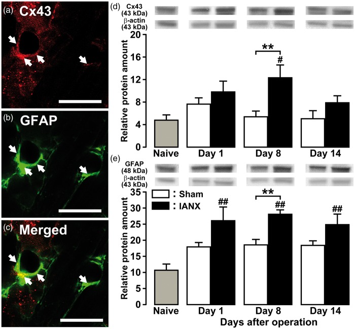
Figure 2.
Cx43 and GFAP expression in TG following IANX. Photomicrographs of Cx43-IR cells (a), GFAP-IR cells (b), Cx43-IR and GFAP-IR cells (c) in TG on day 8 following IANX. Arrows denote double-IR cells. Scale bars: 50 µm. Relative amount of Cx43 (d) and GFAP protein (e) in the TG of naive rats and on days 1, 8, and 14 after IANX or sham operation. β-actin protein was used as loading control. Data represent mean ± SEM. **p < 0.01: compared to sham-operated rats; #p < 0.05, ##p < 0.01: compared to naive rats. (n = 8 in each group; one-way ANOVA followed by Newman-Keuls’s multiple-comparison tests.)

