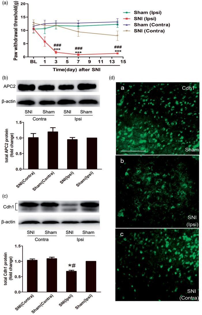
Figure 2.
Changes in APC/C-Cdh1 expression in the dorsal horn after nerve injury. (a) The MWTs of bilateral hind paws to von Frey filament probing were measured, and spared nerve injury (SNI) produced persistent mechanical allodynia in the ipsilateral hind paws starting at day 3 and continuing up to day 14 post-injury (n = 10 in each group). (b and c) Representative bands (top) for the expression of anaphase-promoting complex subunit 2 (APC2) and Cdh1 in the bilateral spinal cord at 14 d after SNI. Quantitative data (bottom) for Western blotting bands (n = 3 in each group) are shown. β-Actin was used as an internal reference, and the fold change for density in the sham (Ipsi) group was set at one for quantification. (d) Immunofluorescence shows Cdh1 expression in the superficial laminae of the dorsal horn at 14 days after SNI. Note that Cdh1 immunoreactivity in the superficial dorsal horn is less intense at the ipsilateral side. Data are expressed as the mean ± standard error of the mean (SEM). ***P < 0.001 versus sham (Ipsi); ###P < 0.001 versus baseline; two-way analysis of variance (ANOVA) followed by Fisher’s least significant difference (LSD) post hoc test. *P < 0.05 versus sham (Ipsi); #P < 0.05 versus SNI (Contra); one-way ANOVA followed by Fisher’s LSD post hoc test. BL: baseline; Contra: contralateral; Ipsi: ipsilateral. Scale bars: 200 µm.

