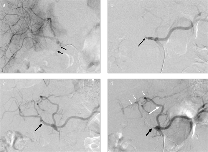
Figure 4.
a–d. A 58-year-old man with hemobilia after the resection of cholangiocarcinoma. Selective angiogram of the common hepatic artery (a) shows extravasation of contrast material through the common hepatic artery (black arrows). Selective angiogram after embolization with two coils (8–50 mm) (b) shows the absence of contrast extravasation and occlusion of the common hepatic artery (black arrow). Because of rebleeding three days after the first TAE, the second selective angiogram (c) shows recanalization of the common hepatic artery (black arrow). Selective angiogram of the celiac artery after embolization with coils (5–50 mm, 8–50 mm) (d) shows occlusion of the common hepatic artery (black arrow) and the accessory left hepatic artery (long white arrow) originating from the left gastric artery, which compensates for the blood supply to the hepatic parenchyma through the communicating vessels (short white arrows).

