
Figure 1.
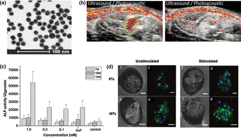
(a) TEM image of 20 nm gold nanospheres.126 (b) In vivo PA/US images of MSCs labeled with 20 nm AuNSs. The left panel is the treatment group, injected with labeled MSCs, and the right panel is the control, no injection.126 (c) Gold nanoparticles can stimulate osteogenic differentiation, as increased AuNP concentrations lead to higher ALP activity.198 (d) Cardiomyocytes seeded in scaffolds with AuNPs and treated with electrical stimulation lead to the highest connexin-43 expression, indicated by green fluorescence. (1, 2, 5, and 6) contain no thiol/(hydroxyethyl)methacrylate (HEMA) whereas (3, 4, 7, and 8) contain 40% thiol/HEMA, providing sites for AuNP growth. The left panel was unstimulated and the right panel was electrically stimulated.201