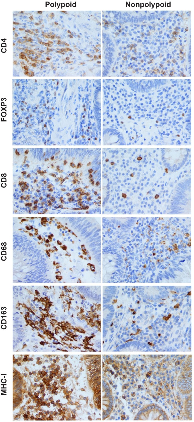
Fig 2. Immunohistochemical assessment of stromal immune cell infiltrates in polypoid and nonpolypoid colorectal adenomas.
CD4+, FOXP3+, CD8+, CD68+, CD163+, and MHC-I+ immune cell densities (i.e., number of positively-stained cells per unit area; see Methods) in the stroma of polypoid adenomas were consistently higher than those observed in nonpolypoid lesions. Intraepithelial CD8+ (cytotoxic) T cells are also seen in the epithelial compartments of both types of tumor. In calculating CD68+ and CD163+ macrophage densities, we counted only nuclei surrounded by CD68+ or CD163+ granulations and excluded elongated or fragmented cells with granular cytoplasmic positivity. MHC-I labelling (granular, compact, or membranous) was observed in a variety of stromal cells. MHC-I+ epithelial cells were also present in both types of tumor, but their presence was not quantified in this study. Magnification for all panels: 400X.

